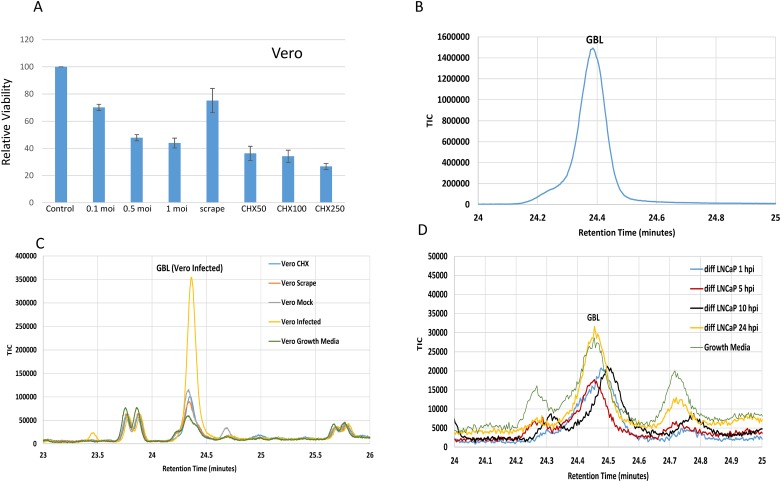
Fig 4. Characterization of GBL induction during stress condition and differentiated LNCaP cells.
A. Relative viability of cells upon infections, scraping, and CHX treatment was evaluated in comparison to control. They were measured at 48 h post infection or post treatment. B. A GBL standard was run as control for GC/MS analysis. The spike was detected approximately at the retention time of 24.4 min. C. GBL was identified only in infected Vero cells but not from CHX, scraping, mock control, and culture media. The CHX concentration was 250 μg/ml. D. The GBL detection was not different in the infected LNCaP cells in comparison to the control. Infection of LNCaP was performed with the same moi of Vero infection.

