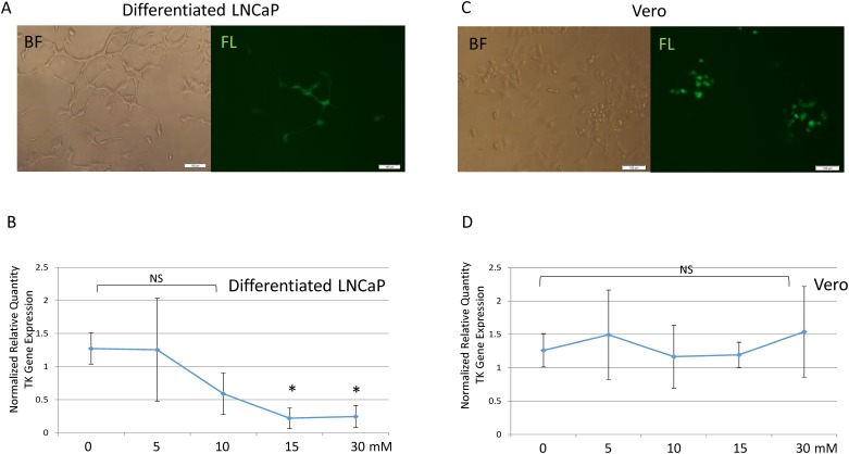
Fig 6. Concentration-dependent effects of GBL on viral gene expression.
A. Fully differentiated LNCaP cells infected by recombinant HSV-1 emitting GFP. The image was captured with an exposure time of 1/6 sec. BF: Bright field; FL: Fluorescent microscopy. B. The concentration-dependent effects of GBL on infection of differentiated LNCaP was determined at 48 hpi by q-RT-PCR measuring HSV-1 TK standardized by the housekeeping gene PPIA. A statistical study was presented using ANOVA with Dunnett’s post-hoc test. Bars labeled with an asterisk (*) were found to be statistically different in comparison to a vehicle with a p<0.05. NS: Not Significant. C. Vero cells were infected by recombinant GFP expressing HSV-1. The exposure time was 1/6 sec when the image was captured. BF: Bright field; FL: Fluorescent microscopy. D. The concentration-dependent effects of GBL on infection of Vero was determined by q-RT-PCR using the same method described in Fig 6B. A statistical study was determined by ANOVA. NS: Not Significant with p>0.05.

