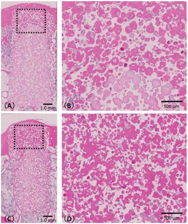
Figure 5. Histological specimen of samples A1 and A2. A1 (A) and A2 (C) are the 24- and 12-week samples, respectively. Newly formed bone was detected in the pores of both A1 and A2. In the center of the cortical bone area (in the dashed box), significantly more bone had been formed in the pores of (D) A1 compared with (B) A2.

