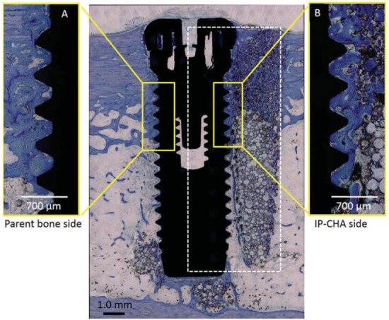
Figure 7. Histological specimen of sample B. (A) High magnification view of the implant surface at the parent bone site. New bone formation from preexisting cortical bone was detected, showing that osseointegration was achieved; (B) High magnification of the implant surface at the IP-CHA site. New bone formed in the IP-CHA pores in contact with the implant surface, showing that osseointegration was achieved. The bottom portion of the IP-CHA pores contained small amounts of new bone. (The dotted line indicates placed IP-CHA).

