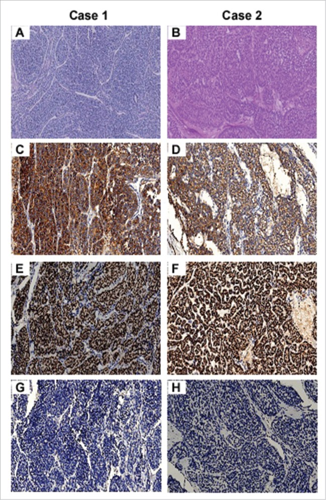
Figure 1.

Histopathological features of two cases of PDTC. Tumor showed typical PDTC growth patterns of insular and trabecular growth in both case 1 (A) and case 2 (B) (hematoxylin-eosin (HE) stain). Immunohistochemical staining for thyroglobulin and thyroid transcription factor 1 (TTF1) is robust in both case1 (C and E) and case 2 (D and F), while staining of calcitonin is absent in both case 1 (G) and 2 (H). All images are 200× magnification.
