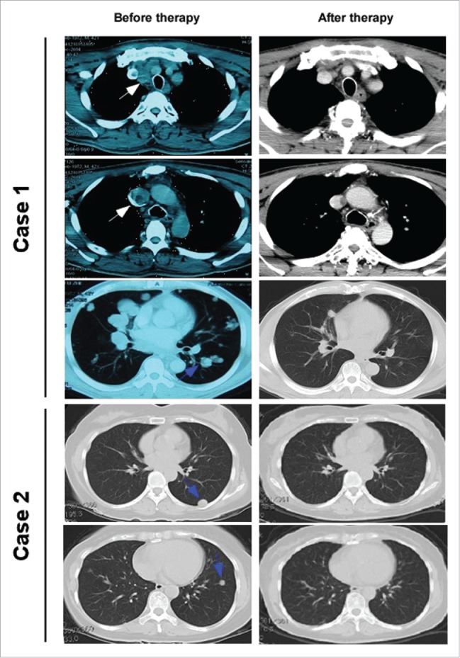
Figure 2.

Chest CT scan images taken prior and subsequent to AD regimen therapy. Enhanced CT of chest with case 1 showed a remarkable reduction in the size of metastatic lymph node located beside trachea and esophagus, and an obvious decrease of cancer embolus (1.2 cm×1.0 cm) in right internal jugular venous (white arrows). Lung window of CT of both case 1and case 2 reveals that a very good response in metastatic lesions of both lungs (blue arrows).
