Figure 6.
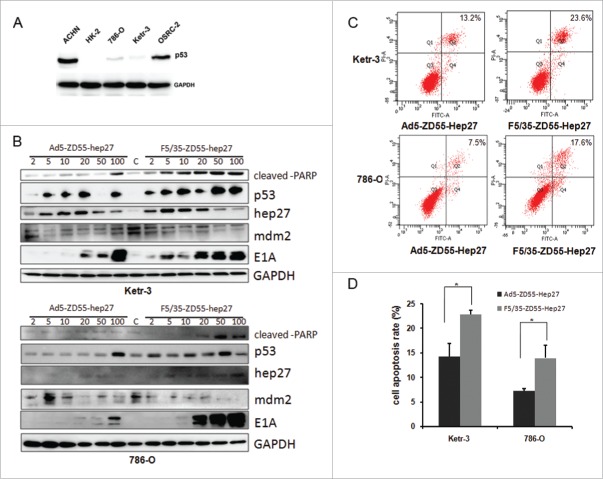
Adenoviral-expressed hep27 inhibited mdm2 and increased p53 and cleaved-PARP expression to induce cell apoptosis. (A) Ketr-3, OSRC-2,786-O, ACHN and HK-2 cells were lysed and lysates were to detect p53 expression by protein gel blotting. GAPDH served as the protein loading control. (B) Ketr-3 and 786-O cells were infected with Ad5-ZD55-Hep27 and F5/35-ZD55-Hep27 at an MOI of 2, 5, 10, 20, 50 and 100 for 48h respectively. Lysates were harvested for western blotting assay. The level of cleaved PARP, p53, mdm2, E1A and hep27 was analyzed. GAPDH served as the protein loading control. (C) Ketr-3 and 786-O cells were infected with Ad5-ZD55-Hep27 and F5/35-ZD55-Hep27 at an MOI of 20 for 48h. The analysis of Annexin V/PI double stained cells was performed to quantify cell apoptosis by FACS. Representative flow cytometric data are shown. (D) The mean percentage of cell apoptosis was calculated based on three independent experiments. * means P < 0.05.

