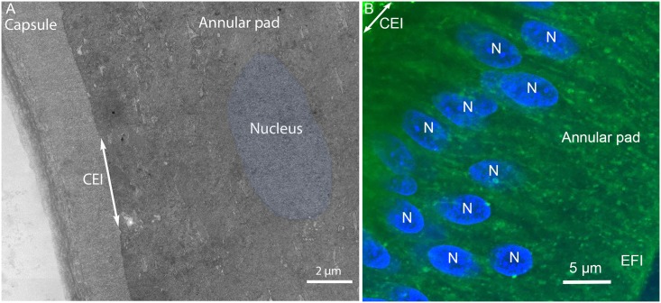
Fig 2. Overview of the capsule and annular pad.
(A) Electron micrograph. (B) Confocal image. The capsule, annular pad and the capsule-epithelial-interface (CEI; double arrow) are indicated. One nucleus (Fig 2A, blue) is highlighted because it is difficult to visualize among the densely stained epithelial cells of the annular pad. Numerous nuclei are stained in the confocal image of an equivalent region of the annular pad (Fig 2B, N). Other membranous organelles such as lysosomes, mitochondria and endoplasmic reticulum are visible as globular objects but not distinguished. EFI is the epithelial to fiber cell interface.

