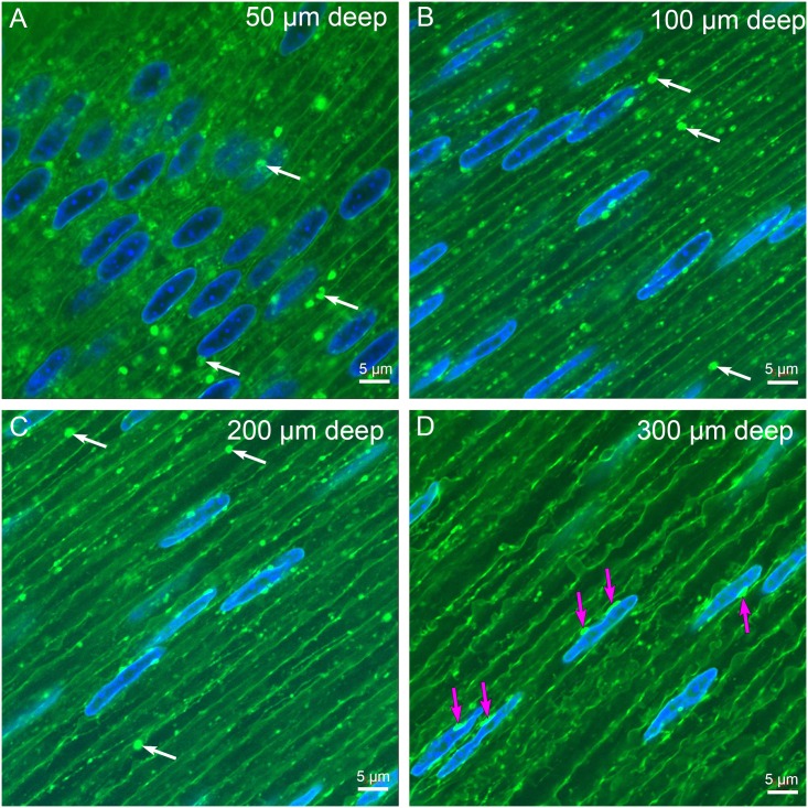
Fig 3. Laser scanning confocal imaging of D15 Vibratome sections.
The depth from the capsule-epithelium-interface is indicated in the upper right corner of each image here and in subsequent images. (A) At 50 μm depth, the nuclei are large, oval and active indicated by the deep 4’,6-Diamidino-2-Phenylindole (DAPI) staining. The cells have uniform parallel borders except where nuclei distort the cellular shape. The membranes stained with 1,1’-Dioctadecyl-3,3,3’,3’-Tetramethylindocarbocyanine Perchlorate (DiI) are uniform in staining and smooth in topology. Numerous vesicular structures are visible that represent membranous organelles including mitochondria, lysosomes, endoplasmic reticulum and Golgi, as well as large circular objects that may represent autophagic vesicles (arrows). (B) At 100 μm depth, the intensely staining vesicular structures (arrows) predominate the background around nuclei that appear to be smaller in average diameter and lighter staining. (C) At 200 μm depth, a few large vesicular structures are visible (arrows), although the overall number of labeled structures is greatly reduced at the outer edge of the organelle-free zone. The nuclei are thin overall and sometimes irregular in width. (D) At 300 μm depth, the nuclei are thin, irregular in width and decorated with oblong brightly staining objects that appear to be attached to the DiI-stained nuclear envelope (magenta arrows). These appear prominent in part because they are not circular and there are very few vesicular structures visible. These structures are the first clear indication that there is a distinct complex that appears to be modifying the nuclei at the edge of the organelle-free zone. Note that the fiber cells are large in diameter and irregular in shape consistent with their depth from the capsule.

