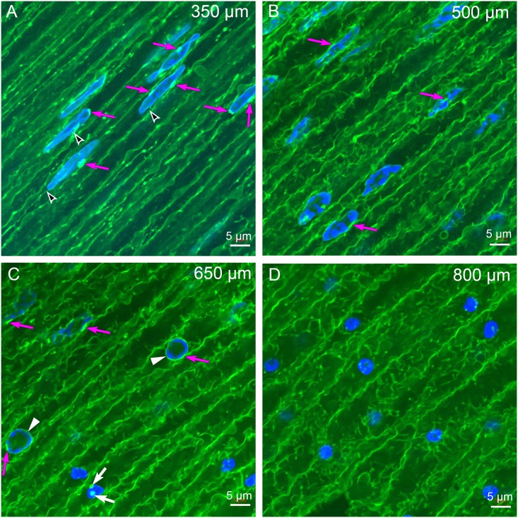
Fig 4. Laser scanning confocal imaging near the organelle-free zone.
(A) At 350 μm depth, the cell shape is irregular and very few vesicular organelles are present within cells. Several examples of the brightly labeled structures located at the nuclear envelope are indicated (magenta arrows), as are potential links to the plasma membrane (black arrowheads). Both these structures can be better appreciated in z-series optical sections (see S1 Fig). (B) At 500 μm depth, the nuclei are irregular in shape and are beginning to degrade. Some nuclei have bright staining close to the nuclear envelope (magenta arrows) while others do not. (C) At 650 μm depth, the cells are larger and more irregular in shape. The nuclei are disrupted with two showing objects associated with the nuclear envelope (magenta arrows, upper left). The two examples in the center have circular shapes, faint staining of associated structures (magenta arrows) and light staining of the nuclear envelope (white arrowheads). These appear to be in the final stages of breakdown of the nuclear envelope as the four examples on the lower right are small globular remnants of nuclei with no visible nuclear envelope. Two small circular dots (white arrows) may be remnants of nuclear envelope breakdown but are not consistent with the vesicular structures in previous images. (D) At 800 μm depth, the cells are very large and irregular with several globular remnants of nuclear breakdown stained brightly with 4,6-diamidino-2-phenylindole (DAPI). These are most likely the pyknotic nuclei described in the early literature.

