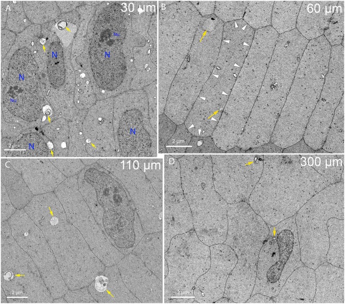
Fig 5. Fiber cell ultrastructure near the bow region up to a depth of 300 μm.
(A) Nuclei of the bow region are present among well-defined radial cell columns. Many autophagic vesicles (arrows) are visible as well as numerous other smaller organelles. Two nuclei (N) have clear nucleoli (Nu). Day 15 lens imaged 30 μm from the capsule-fiber cell interface. (B) Just beyond the nuclei of the bow region, classical fiber cells with hexagonal 2 μm x 10 μm cross-sections in radial cell columns are observed. Autophagic vesicles (arrows) are present as are other organelles where protein production is still occurring. The dark stained globular clusters in the cytoplasm are probably polysomes. Extensive gap junctions that appear as thin dark lines on broad and narrow faces cover about 50% of the cell surface as indicated for one cell (arrowheads). Day 15 lens about 60 μm from the capsule-fiber cell interface. (C) Fiber cells just outside the organelle-free zone (OFZ) appear normal. Note the nucleus is slightly irregular in shape but otherwise normal in appearance with portions of a nucleolus visible. Autophagosomes with double membranes are indicated (yellow arrows), although they decrease in number corresponding to a decrease in membranous organelles in the cytoplasm through this region. The fiber cells have hexagonal shapes, which are somewhat irregular in size and slightly more rounded. Day 15 lens about 110 μm from the capsule-fiber cell interface. (D) Nuclei at the edge of the OFZ display irregular shapes. Note the pronounced indentation and thin region, which if projected along the nuclear length would suggest a significant reduction in nuclear volume. Irregular nuclear shapes including indentations have been reported previously [29]. Autophagic vesicles (arrows) and other organelles including mitochondria and endoplasmic reticulum are also present although quite rare. An important feature of this region is the irregular shape and rounded edges of the fiber cells. Day 15 lens about 300 μm from the capsule-fiber cell interface.

