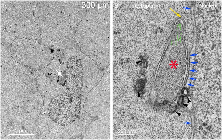
Fig 6. Indentations often reveal a large macromolecular complex adherent to the nuclear envelope.
(A) Low magnification shows a complex (arrow) that has an unusual asymmetric shape and irregular densities associated with one surface. (B) High magnification is necessary to identify the components of the complex. The core (asterisk) has a uniform texture similar to the adjacent cytoplasm (distinct from nucleoplasm, lysosomes or any type of autophagosome) and is surrounded mainly by two membranes. The outer membrane is closely adhered to the outer nuclear envelope and distorting that membrane (five central blue arrows). The other segments of the outer nuclear membrane (remaining blue arrows) have a normal association with the inner nuclear membrane. The tip is narrowed and covered by a protein layer (yellow arrow) that is 25 nm thick with repeating protein densities that are similar to clathrin and its adapter proteins. The interior of the tip contains fibers (green lines) that are similar actin microfilaments often seen in lens epithelium, fiber cell cytoplasm and elongating ball-and-socket devices. The dense bodies (arrowheads) are multilayered bilayers with 5 nm spacing typical of pure lipid (without integral proteins). Day 15 lens 300 μm from the capsule-fiber cell interface.

