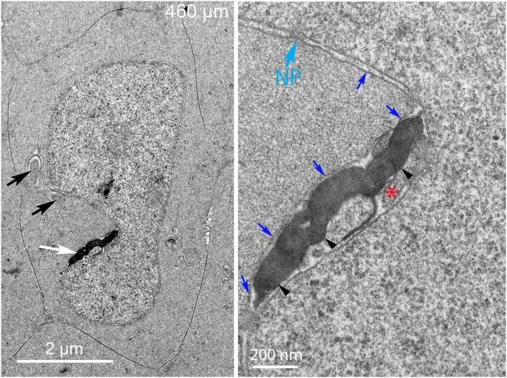
Fig 9. Many complexes contain large aggregates of thin multilamellar membranes.
(A) Low magnification shows a large complex in a nuclear indentation with an intensely stained aggregate within the complex (white arrow). In addition, there are two extended objects adjacent to the nucleus (black arrows) that are early stages of complex formation as discussed below. (B) High magnification shows that the large dark object is an extensive collection of thin bilayer membranes (arrowheads) that most likely represent pure lipid derived from the breakdown of the nuclear envelope. Bilayer average spacing was 5.2 nm (n = 59). The complex is contained beneath the outer nuclear membrane (blue arrows). The presumed core (asterisk) is small but similar in texture to the adjacent cytoplasm and not to autophagosomes or lysosomes. An intact nuclear pore (NP) is indicated. Day 15 lens 460 μm from the capsule-fiber cell interface.

