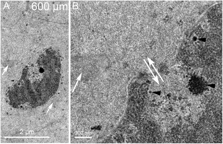
Fig 13. Nuclear envelope breakdown in a localized region reveals multiple staining patterns of the nucleoplasm.
(A) Low magnification shows two projections near the nucleus (arrows) that exhibits several light staining regions as if material has been removed. (B) High magnification of a light-staining region shows that the nuclear envelope has been lost (double arrows) and that the nucleoplasm aggregates into small and large particles (arrowheads), some of which may have departed the nucleoplasm to create the lighter staining zones. This structure may represent a nucleus just prior to becoming pyknotic near the organelle-free zone. Day 15 lens about 600 μm from the capsule-fiber cell interface.

