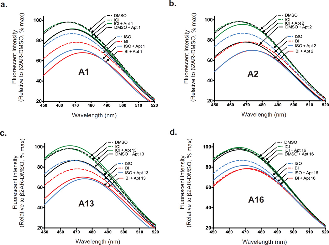
Figure 4. Influence of aptamers on conformational changes conferred by ligands.
(a–d) Bimane fluorescence quenching measurement shows that aptamers A1, A2, and A13 stabilize active forms of β2AR, while aptamer A16 stabilizes an inactive conformation. Bimane fluorescence quenching measurement detects conformational changes of the β2AR via movement of a bimane probe on TM6 (at C265) upon the binding of agonists, (BI167107 [BI] or isoproterenol [ISO]) and/or aptamer: A1 (a), A2 (b), A13 (c) or A16 (d). Fluorescence emission spectra showing ligand-induced conformational changes of bimane-labeled β2AR in the absence (black dashed line) or presence of full agonist (ISO, blue dashed line, or BI, red dashed line), inverse agonist ICI-118,551 (ICI, green dashed line), aptamer A1, A2, A13, or A16 (black solid line), or a combination of aptamer (A1, A2, A13, or A16) with ISO (blue solid line), BI (red solid line), or ICI (green solid line).

