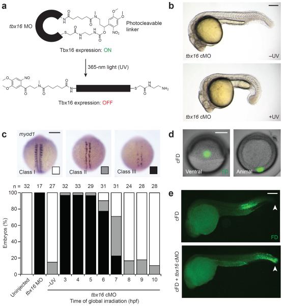
Figure 1. Optochemical regulation of Tbx16-dependent paraxial mesoderm development.
(a) Schematic representation of a cyclic tbx16 cMO and its photoactivation. (b) Phenotypes observed in embryos injected with the tbx16 cMO and cultured in the dark (−UV) or globally irradiated at 3 hpf (+UV). (c) Phenotypic classes of myod1 expression observed at 13 hpf and the distributions associated with global irradiation of tbx16 cMO-injected embryos at the designated times. (d) Live embryos injected with cFD and irradiated within the ventral margin at 6 hpf to target trunk somite progenitors. (e) Immunostaining of uncaged fluorescein in fixed 24-hpf embryos that were previously injected with cFD alone or a cFD/tbx16 cMO mixture and then irradiated as described for panel d. White arrowheads indicate the tailbud. Embryo orientations: a–b and e, lateral view, anterior left; c, dorsal view, anterior up; d, ventral and animal pole views. Scale bars: 200 μm.

