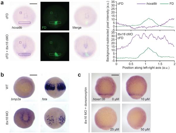
Figure 4. Non-cell-autonomous signals propagate posterior hox gene activation in Tbx16-deficient embryos.
(a) hoxa9b expression in 10-hpf zebrafish embryos that were previously injected with cFD alone or a cFD/tbx16 cMO mixture and irradiated within the ventral margin at 6 hpf. Contrast-enhanced micrographs are shown (left), and unprocessed images were used to determine integrated pixel-intensity profiles (right) across the indicated regions (dashed rectangles) and background signals (dashed squares). Note that the hoxa9b expression domain in cFD/tbx16 cMO-injected embryos extends beyond the irradiated cells, which are labeled with anti-fluorescein antibodies. (b) Staining of bmp2a transcripts in the ventral lateral margin and fsta transcripts in the anterior paraxial mesoderm of wild type and tbx16 morphant embryos at 9 hpf. (c) hoxa13b expression in 8-hpf tbx16 morphants that were treated with the indicated doses of dorsomorphin from 4 hpf onward. In comparison, wild type embryos do not express hoxa13b at this stage (see Fig. 3a). Embryo orientations: a, vegetal pole view, dorsal up; b, bmp2a, vegetal pole view and dorsal up; fsta, dorsal view and animal pole up; c, ventral view, animal pole up. Scale bars: 200 μm.

