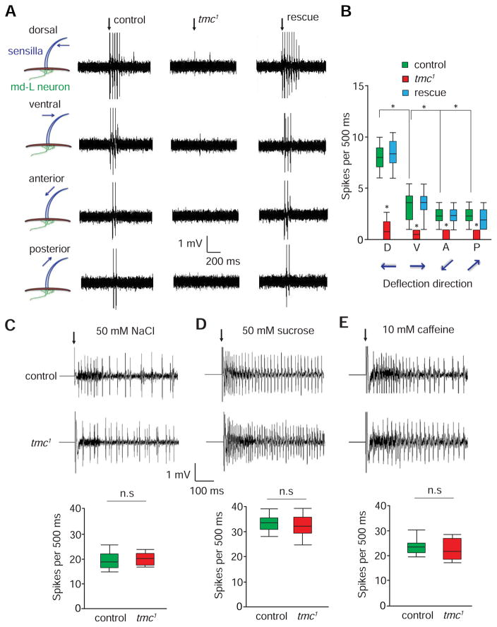
Figure 4. Electrophysiological responses of md-L neurons to mechanical stimuli.
(A) Bending of L-type taste bristles exerted compression forces underneath dendritic arbors of md-L neurons, thereby leading to the firing of action potentials in md-L neurons. L4 taste bristles from the indicated flies were deflected 20 μm in the dorsal, ventral, anterior and posterior directions. The rescue flies were tmc-Gal4/UAS-tmc;tmc1. The vertical arrows indicate the onset of the mechanical stimuli.
(B) Box and whisker plots showing the number of spikes/500 ms. n=10.
(C—E) Tip recording traces showing the responses of control and tmc1 GRNs to chemical stimulation. The box and whisker plots show the summary tip recording data. Not significant, n.s. n=5.
(C) 50 mM NaCl (L4 sensilla).
(D) 50 mM sucrose (L4 sensilla).
(E) 10 mM caffeine (S6 sensilla).
The error bars indicate SEMs. *p< 0.05. ANOVA tests with Scheffé’s post-hoc analysis.

