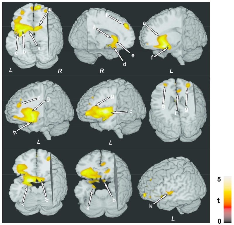
Figure 2.
Negative correlations between MTR values and AHI scores in OSA subjects. Brain sites included the caudate nuclei (a), putamen, extending to internal and external capsules (b), left insular cortex (c), ventral medial prefrontal cortex and surrounding white matter (d), genu of corpus callosum (e), bilateral superior frontal cortices, extending to the surrounding white matter (f), anterior cingulate and cingulum bundle (i), prefrontal cortices (g), hypothalamus (f), amygdala and hippocampus (j), left ventral temporal lobe (k), inferior frontal cortex, and surrounding white matter (h). All images are in neurological convention (L = Left; R = Right). Color bar indicates t-statistic values.

