
Figure 2.
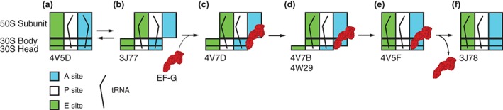
Scheme of tRNA rearrangements during EF‐G‐catalyzed ribosome translocation. Diagrams show tRNA positions relative to the A, P, and E sites on the 50S subunit and 30S head and body. (a) peptidyl‐ and deacylated tRNAs are bound in A/A and P/P classical states, (b) A/P and P/E hybrid states, (c) A/P* and P/E states in the presence of ribosome‐bound EF‐G, (d) ap/P and pe/E chimeric states in the presence of ribosome‐bound EF‐G, (e) classical P/P and E/E state in the presence of ribosome‐bound EF‐G, and (f) classical P/P and E/E state after EF‐G dissociation. Please see additional details in the text. PDB IDs corresponding to each structural state are indicated under the schematic.