
Figure 4.
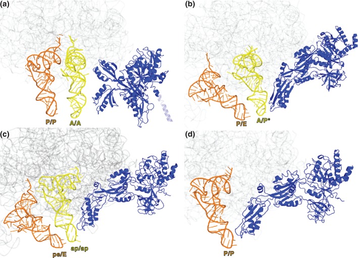
Structural rearrangements of EF‐G on the ribosome during translocation. (a) EF‐G (blue in all structures) bound to the nonrotated pretranslocation ribosome containing A/A‐ and P/P‐ tRNAs93 (PDBID 4WPO). The N‐terminal domain of large subunit protein L9 covalently linked to the N‐terminus of EF‐G is shown as a transparent blue. (b) EF‐G bound to the fully rotated pretranslocation ribosome containing A/P*‐ and P/E‐ tRNAs55 (PDBID 4V7D); (c) EF‐G bound to partially rotated ribosomes containing chimeric ap/ap and pe/E‐ tRNAs42 (PDBID 4 W29) and (d) EF‐G bound to the nonrotated posttranslocation ribosome containing P/P‐ and E/E‐ tRNAs13 (PDBID 4V5F). 23S rRNA in all structures is shown in gray.