
Figure 6.
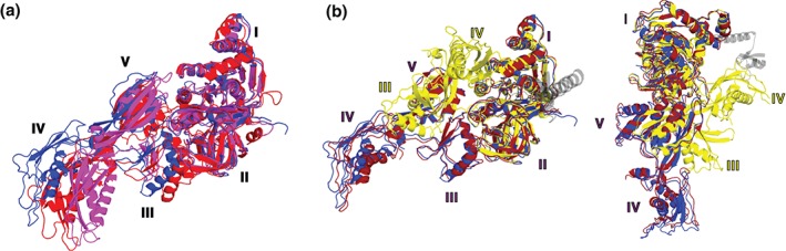
Interdomain rearrangements of EF‐G. (a) EF‐G in the posttranslocation conformation (blue, PDBID 4V5F13) is superimposed by structural alignment of domains I and II with ribosome‐free EF‐G•GDP (magenta, PDBID 1DAR2) and EF‐G bound to the viomycin‐trapped pretranslocation ribosome (red, PDBID 4V7D55), (b) EF‐G in the posttranslocation conformation (blue, PDBID 4V5F13) is superimposed by structural alignment of domains I and II with EF‐G bound to a ribosome containing a chimeric ap/ap tRNA (dark red, PDBID 4W2942) and EF‐G‐L9 fusion bound to the nonrotated, pretranslocation ribosome (yellow, PDBID 4WPO93). The N‐terminal domain of L9 covalently linked to EF‐G (PDBID 4WPO93) is shown in transparent gray. Two differently oriented views of EF‐G structures are shown. Domains of EF‐G are numbered as indicated.