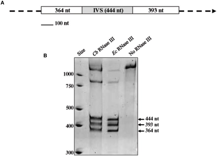
Figure 2.
In vitro processing of the IVS by RNase III. (A) Map of the RNA substrate used in RNase III assays, consisting of a segment of 23S rRNA with the IVS (gray box) and proximal flanking sequences (white boxes) with sizes (in nucleotides) shown. (B) Acridine orange-stained polyacrylamide gel [4% acrylamide (w/v)—8M urea] showing cleavage products of substrate RNA following treatment with recombinant C. burnetii (Cb) RNase III. Three discrete fragments of sizes corresponding to the substrate RNA (arrowed) were observed. Positive [E. coli (Ec) RNase III (Ambion)] and negative (no RNase III) control reactions are shown for comparison.

