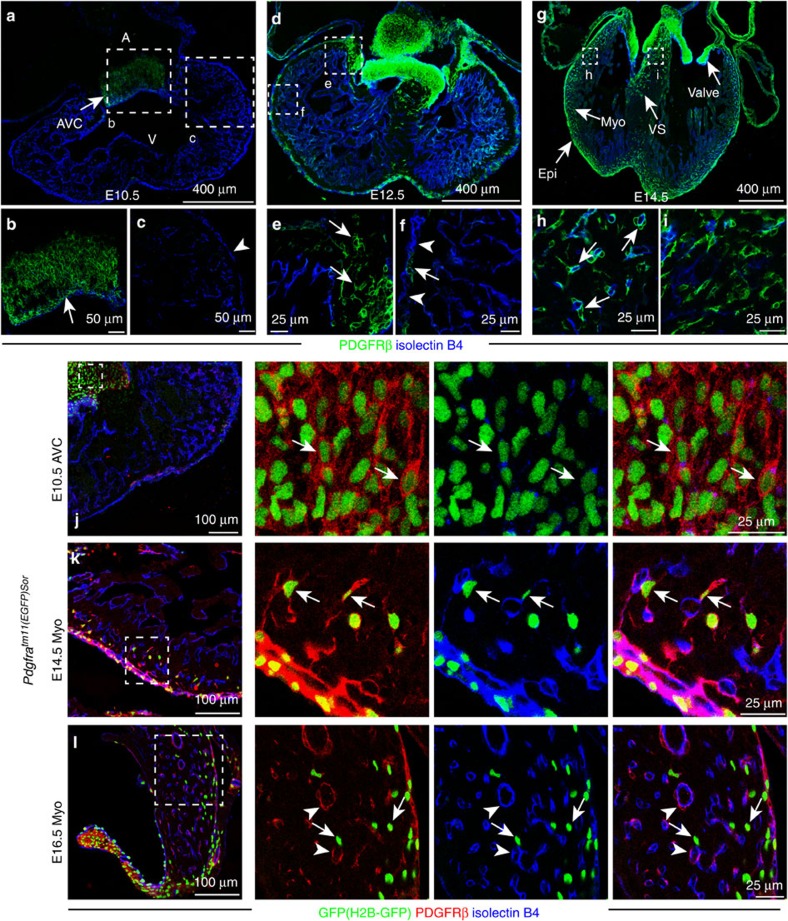
Figure 1. Developmental distribution and molecular properties of PDGFRβ+ cells.
(a–i) Distribution of PDGFRβ (green) immunostained cells from E10.5 to E14.5. Heart sections from wild-type mice were stained for PDGFRβ (green) and isolectinB4 (blue). Arrows indicate PDGFRβ+ cells, arrowheads mark PDGFRβ- epicardial cells at the indicated stages. Panels at the bottom (b,c,e,f,h,i) are higher magnifications of insets in (a,d,g), respectively. A, atrium; V, ventricle; AVC, atrioventricular canal; Epi, epicardium; Myo, myocardium; VS, ventricular septum. (j–l) Transient co-expression of PDGFRβ (red) and PDGFRα (Pdgfratm11(EGFP)Sor reporter; nuclear H2B-GFP; green) during heart development. ECs, isolectinB4 (blue). Arrows indicate GFP+ PDGFRβ+ double-positive cells in the E10.5 AVC (j) and E14.5 myocardium (k). Double-positive cells were very rare in the E16.5 myocardium (l), whereas GFP- PDGFRβ+ mural cells (arrowheads) and GFP+ PDGFRβ- interstitial cells (arrows in l) were abundant.

