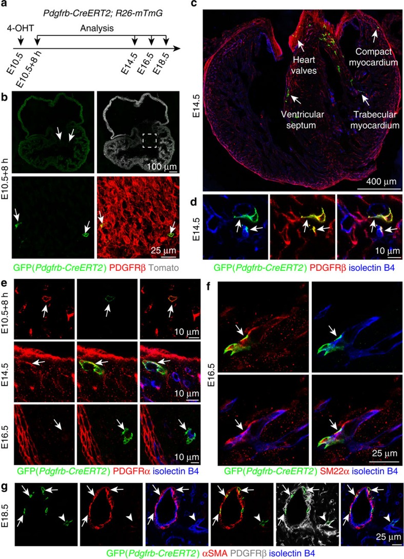
Figure 2. Cardiac mural cells originate from mesenchymal progenitors.
(a–g) Clonal analysis using Pdgfrb(BAC)-CreERT2 Rosa26-mTmG mice. (a) Experimental strategy indicating the stages of 4-hydroxytamoxifen (4-OHT) administration and analysis. (b) GFP+ cells (green, arrows) in AVC at 8 hours after 4-OHT administration. PDGFRβ (red) immunostaining and unrecombined cells (tdTomato, white/red) are shown. (c,d) Overview of GFP+ cell distribution in the indicated regions of E14.5 heart (c). At higher magnification, GFP+ PDGFRβ+ cells (arrows) in the myocardium were found in association with isolectin B4-labelled vessels (blue) (d). (e) Arrows indicate representative GFP+ cells, which were PDGFRα+ (red) in the E10.5 AVC and E14.5 myocardium, but PDGFRα- and located at isolectin B4+ vessels (blue) in E16.5 myocardium. (f) Pdgfrb(BAC)-CreERT2-labelled cell clones (GFP, green) gave rise to SM22α+ mural cells (red, arrow) at E16.5. ECs, isolectin B4 (blue). (g) Pdgfrb(BAC)-CreERT2-marked cell clones (GFP, green) were identified as αSMA+ vSMCs (red, arrows) at E18.5. Arrowheads mark a αSMA- perivascular cells. ECs, isolectin B4 (blue).

