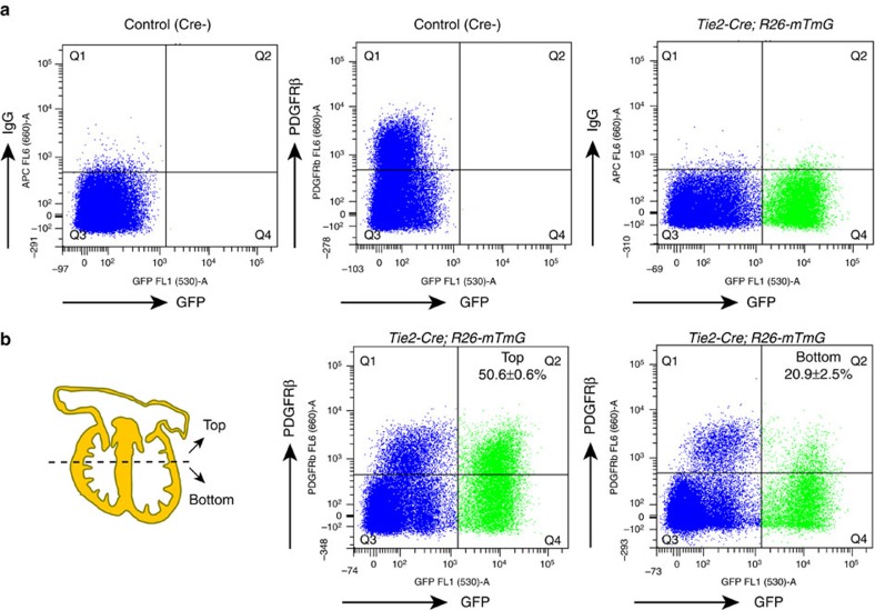
Figure 4. Contribution of endothelial cells to cardiac mural cells.
(a,b) Flow cytometry analysis of cardiac PDGFRβ+ cells of endothelial cell origin at E14.5. (a) Littermate Tie2-Cre-negative mice, IgG-APC and PDGFRβ-APC antibodies were used to define the gating of signals. (b) The top and bottom of hearts were analysed independently. Numbers indicate percentage of GFP+ PDGFRβ+ cells relative to total PDGFRβ+ population in top or bottom parts of heart, respectively. n=3 (4 or 5 hearts/experiment). Error bars±s.e.m.

