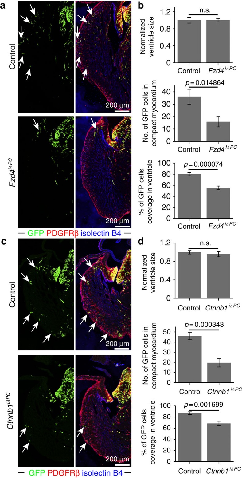
Figure 6. PDGFRβ+ cells require Wnt signalling for recruitment to the compact myocardium.
(a) Maximum intensity projections showing the distribution of Pdgfrb(BAC)-CreERT2-labelled GFP+ cells (green; arrows) in Fzd4iΔPC mutant and littermate control (Pdgfrb(BAC)-CreERT2+/T Fzd4lox/+) E14.5 heart sections. Note profound reduction of GFP+ cells in Fzd4iΔPC compact myocardium. (b) Statistical analysis of Fzd4iΔPC versus control ventricle size, number of GFP+ cells in E14.5 compact myocardium and relative coverage of ventricle by GFP+ cells (n=8 in each group). Error bars,±s.e.m. P values, two-tailed unpaired t-test; NS, no significance. (c) Maximum intensity projections showing Pdgfrb(BAC)-CreERT2-labelled, GFP+ cells (green; arrows) in Ctnnb1iΔPC mutant and littermate control (Pdgfrb(BAC)- CreERT2+/T Ctnnb1lox/+) E14.5 heart sections. GFP+ cells showed reduced migration to the apex and were less abundant in the Ctnnb1iΔPC compact myocardium. (d) Statistical analysis of Ctnnb1iΔPC versus control ventricle size, number of GFP+ cells in E14.5 compact myocardium and relative coverage of ventricle by GFP+ cells (Ctnnb1iΔPC: n=7; control: n=8). Error bars±s.e.m. P values, two-tailed unpaired t-test; NS, no significance.

