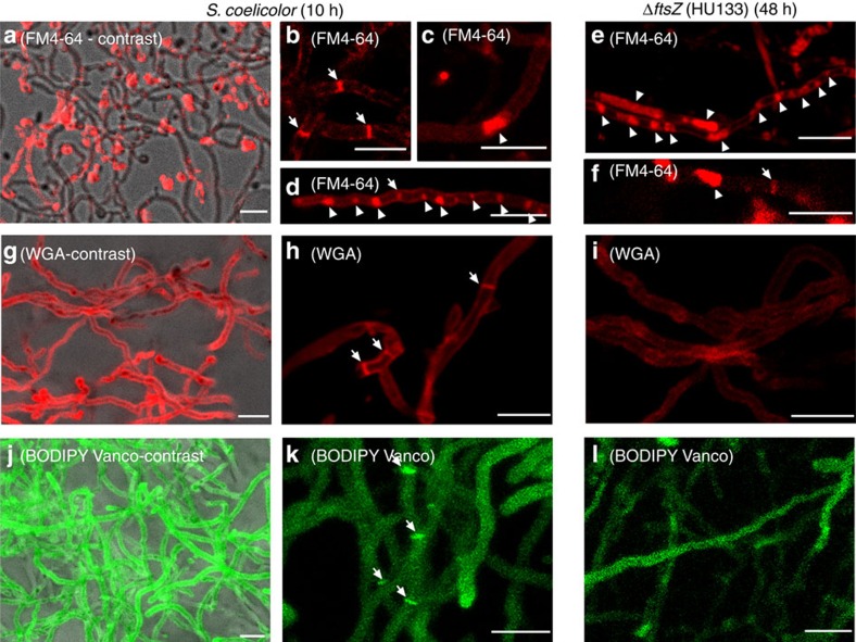
Figure 6. Membrane and cell wall staining of S. coelicolor and its ftsZ mutant HU133.
(a–f) FM4-64 staining (membranes). (g–i) WGA staining (cell wall). (j–l) BODIPY-vancomycin staining (nascent peptidoglycan). Fluorescent images in a, g and j correspond to the maximum projection 10-μm series overlaid with their respective phase-contrast images, showing 100% of the stained hyphae. Arrows indicate cross-membranes and cross-cell walls. Arrowheads indicate membrane cellular segments filled with membrane vesicles. Scale bars, 4 μm.

