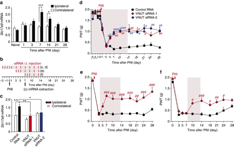
Figure 3. PNI-induced upregulation of VNUT in the spinal cord is required for the development of tactile allodynia.
(a) Real-time PCR analysis of Slc17a9 mRNA in total RNA extracted from spinal cord ipsilateral and contralateral to PNI before (naive) and after PNI. Values represent the relative ratio of Slc17a9 mRNA (normalized to the value for 18S mRNA) to the contralateral side of naive mice (n=6; *P<0.05, ***P<0.001, two-way ANOVA with post hoc Bonferroni test). (b) Experimental schedule. (c) Real-time PCR analysis of Slc17a9 mRNA in the spinal cords of mice treated with control or VNUT siRNAs (20 pmol per injection) 7 days after PNI. Values represent the relative ratio of Slc17a9 mRNA (normalized to the value for 18S mRNA) to the contralateral side of mice with control RNA (control RNA: n=8, VNUT siRNA-1: n=5, VNUT siRNA-2: n=5; *P<0.05, **P<0.01, ***P<0.001, one-way ANOVA with post hoc Tukey Multiple Comparison test). (d–f) Reversal of PNI-induced tactile allodynia by intrathecal administration of VNUT siRNA (20 pmol per injection) in the ipsilateral side of WT mice (d, control RNA: n=5, VNUT siRNA-1: n=4, VNUT siRNA-2: n=5; e, n=5 per group; f, control RNA: n=4, VNUT siRNA-1: n=5; #P<0.05, ##P<0.01, ###P<0.001 versus control RNA, two-way ANOVA with post hoc Bonferroni test). Values are means±s.e.m.

