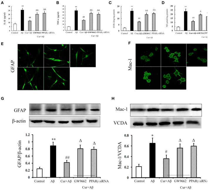
Figure 5.
Curcumin inhibited neuroinflammation in mixed neuron/glia cultures. Mixed neuron/glia cultures were pre-treated with curcumin 10 μM, 1 h later, Aβ1–42 25 μM was added to the mixed cultures. GW9662 1 μM was added into the cultures or cells were transfected with PPARγ siRNA 1 h before Aβ1–42 treatment. (A) IL-1β level of mixed neuron/glia cultures. (B) TNF-α level of mixed neuron/glia cultures. (C) COX-2 level of mixed neuron/glia cultures. (D) NO level of mixed neuron/glia cultures. Data were expressed as mean ± SD with six individual experiments. (E) Immunofluorescence of GFAP. (F) Immunofluorescence of Mac-1. Representative images from five experiments were shown. (G) Western blot of GFAP in mixed neuron/glia cultures. (H) Western blot of Iba-1 in mixed neuron/glia cultures. A representative immunoblot from four independent experiments was shown. Data were expressed as mean ± SD. *P < 0.05, **P < 0.01 vs. control cells, #P < 0.05, ##P < 0.01 vs. Aβ1–42-challenged cells, ΔP < 0.05, ΔΔP < 0.01 vs. curcumin treated cells.

