Figure 2.
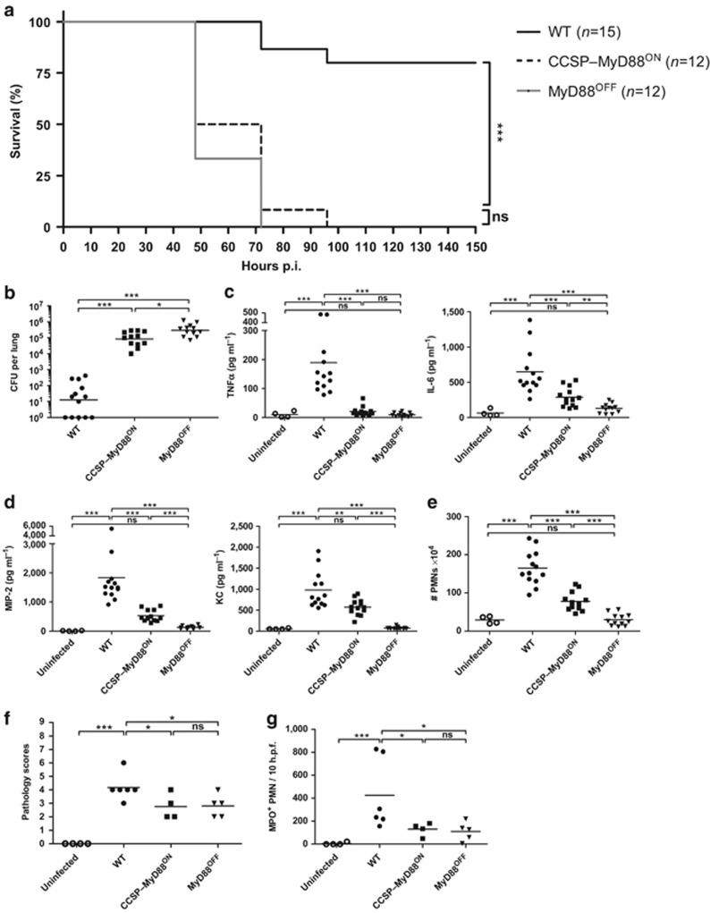
Myeloid differentiation factor 88 (MyD88) signaling in club cells contributes to proinflammatory cytokine production and control of bacterial burden. Wild type (WT), club cell secretory protein (CCSP)–MyD88ON and MyD88OFF mice were intranasally inoculated with either a low dose (a) or a high dose (b–g) of Streptococcus pneumoniae. (a) Survival curve of S. pneumoniae–infected mice. Data show two pooled individual experiments with five to nine mice per group. Statistics were calculated using log-rank (Mantel-Cox) test; ***P<0.001 and ns, not significant. (b) S. pneumoniae burden in total lung homogenates 18 h post infection (p.i.). (c) Tumor necrosis factor α (TNFα), interleukin-6 (IL-6), and (d) macrophage inflammatory protein 2 (MIP-2) and keratinocyte chemoattractant (KC) levels in total lung homogenates 18 h p.i. measured by enzyme-linked immunosorbent assay (ELISA). (e) Fluorescence-activated cell sorting (FACS) analysis of CD11c− MHCII− CD11bhigh Ly6Cint neutrophils (PMNs; excluding CD11c− MHCII− CD11bint Ly6Chi monocytes). Shown are total cell numbers in lungs from WT, CCSP–MyD88ON, and MyD88OFF mice 18 h p.i. Evaluation of (f) hematoxylin and eosin (H&E) and (g) myeloperoxidase (MPO) staining of lung tissue from uninfected and S. pneumoniae–infected mice 18 h p.i. Tissue sections were evaluated in a blinded manner. Dot plots depict (f) histopathological scoring and (g) MPO+ PMNs counted within 10 high power fields (h.p.f.). (b–e) Data represent three pooled individual experiments with three to five mice per group. (f,g) Data are pooled from two individual experiments with two to three mice per group. (b–g) Data points depict individual mice. Bars indicate mean value. Statistics were calculated using Mann-Whitney test; *P<0.05, **P<0.01, and ***P<0.001.

