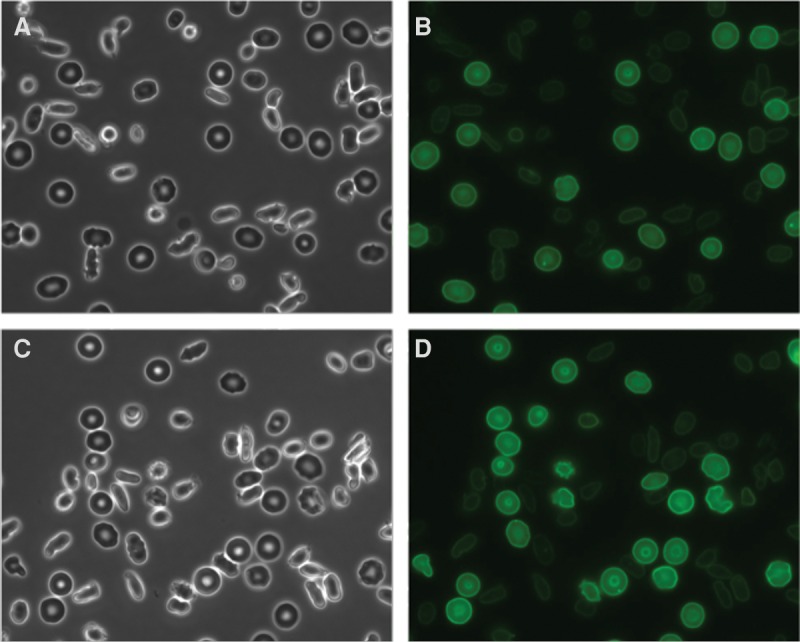
Figure 2.

Microscopic images of representative fields of red blood cells (RBCs) from the EPB41-deficient patient. (A,C) Phase-contrast and (B,D) corresponding fluorescent images of Alexa 488 anti-GPC labeled RBCs. The images were taken using a fluorescent microscope (Nikon Ti) with a 60× oil immersion objective.
