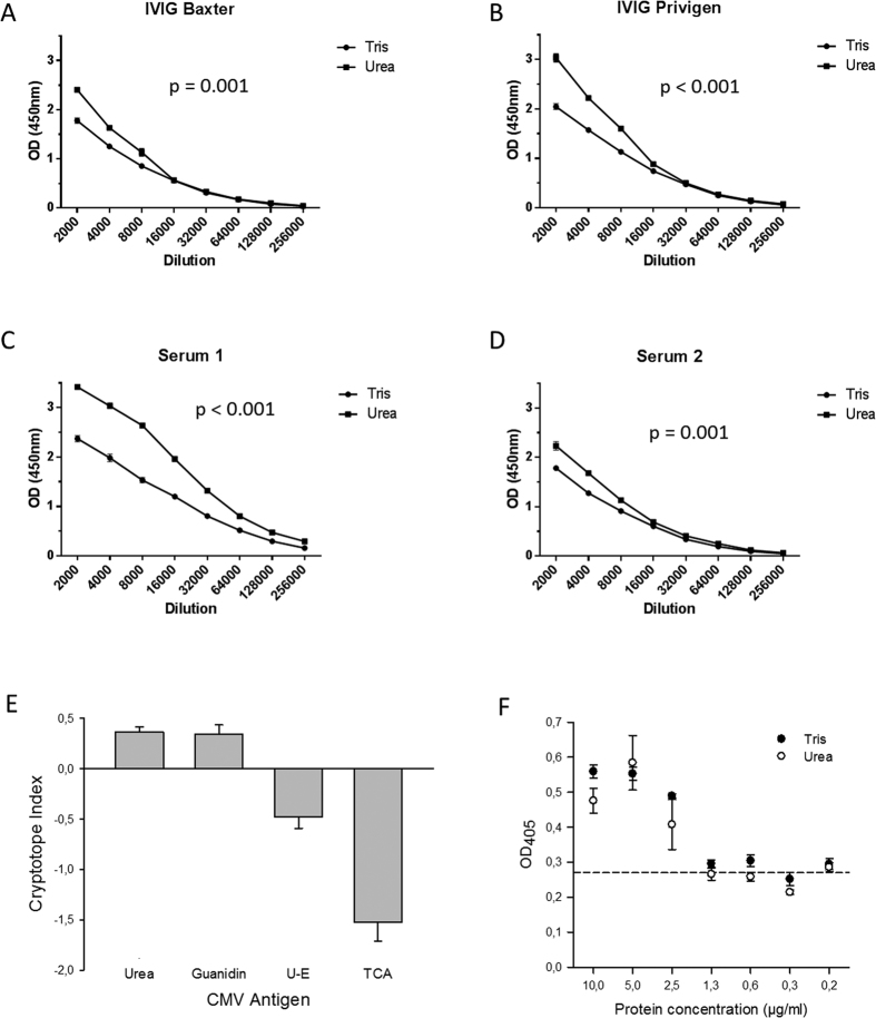
Figure 1. ELISA analysis of the reactivity of human polyclonal immune serum with native and denatured whole CMV lysate.
(A–D) Representative OD450 readings obtained when probing native whole CMV lysates (Tris-buffer) or unfolded whole CMV lysates (Urea-buffer) with two-fold serial dilutions of immunoglobulin preparations (IVIG) and human polyclonal immune serum; error bars indicate standard error or the mean (SEM). (E) Mean OD450 readings obtained when probing native (Tris-buffer), unfolded (Urea- or Guanidin-buffer), or precipitated whole CMV lysate (ethanol (U-E) or TCA) with human polyclonal immune sera at a standard dilution (1:500; n = 18). Bars indicate mean CI measured and error bars indicate the standard error of the mean (SEM). (F) ELISA plates were coated with BSA preparations at different final concentrations, washed thoroughly, and stained with a silver stain to visualize protein adsorbed to the plates. The dashed line indicated mean OD405 readings measured for blanks. Optical density was measured at 405 nm and not corrected for reference measurements at 620 nm because silver absorbs light at both wave-lengths. All BSA experiments were done in triplicates.

