
Figure 7. P. gingivalis LPS1690-induced TXN1 and MnSOD (SOD2) protein expression in HGFs.
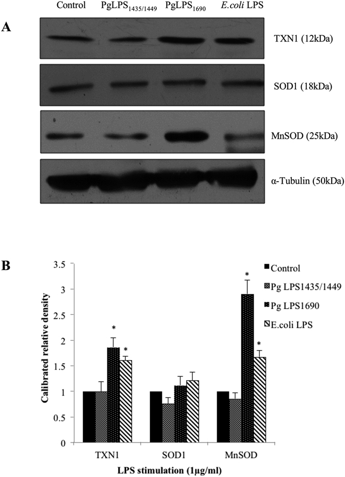
HGFs were treated with P. gingivalis LPS and E. coli LPS at 1 μg/ml for 24 h. Protein was pooled from triplicate samples and 40 μg aliquots of total protein extracts were subjected to Western blot analysis. The membranes were probed with rabbit anti-TXN1 mAbs (1:1000), rabbit anti-SOD1 abs (1:1000) and rabbit anti-SOD2/MnSOD mAbs (1:1000) (A). For loading control, the membrane was stripped again and incubated with rabbit anti-α-Tubulin mAbs (1:2000). The fold increase values of proteins as compared with α-Tubulin are shown in the graphs (arbitrary units over control after normalization to the total protein) (B). One representative blot was shown from three independent experiments with similar results. *Significant difference with a P-value <0.05 as compared with the controls without LPS treatment.