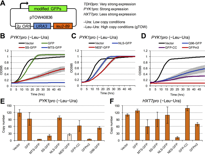
Figure 1. High-level expression of modified GFPs affects cellular growth.
(A) Structure of the plasmid used in this study. Modified GFPs are expressed from TDH3pro, PYK1pro, or HXT7pro. The copy number of the plasmid is low under –Ura conditions and high under –Leu–Ura conditions, owing to the selection bias imposed by leu2d (for details of the gTOW experiment, see Supplementary Method). (B–D) Growth curve of cells expressing modified GFP under –Leu–Ura conditions. The optical densities at 595 nm (OD595) of the cell cultures were measured every 30 minutes. Error bars represent standard deviation. (E,F) Copy numbers of the gTOW plasmids containing modified GFPs under –Leu–Ura conditions. Modified GFPs were expressed from PYK1pro (E) or HXT7pro (F). Error bars represent standard deviations.

