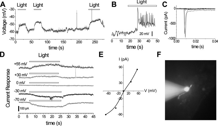
Figure 3.
A. Light-evoked responses from a neuron in the lateral septal organ, showing slowly rising subthreshold depolarization under current clamp. B. Light-evoked response from another cell under current clamp, showing a light-evoked slow depolarization, which led to delayed suprathreshold spikes. C. Voltage-gated Na currents (shown after leak subtraction) from the same cell as in B, in response to a series of depolarizing voltage steps of increasing amplitude from a holding potential of −70 mV. D. Light-evoked current responses recorded at various holding potentials under voltage clamp, showing a reversal potential near 0 mV. E. Current: voltage (I-V) relation of light evoked currents in D. F. Digital image of an Alexa Fluor 594-filled neuron near the ventricular surface of a septal slice under whole-cell patch showing a bipolar morphology. The intensity of full-field light stimulation was 0.04 nW µm−2. Color version available in the online PDF.

