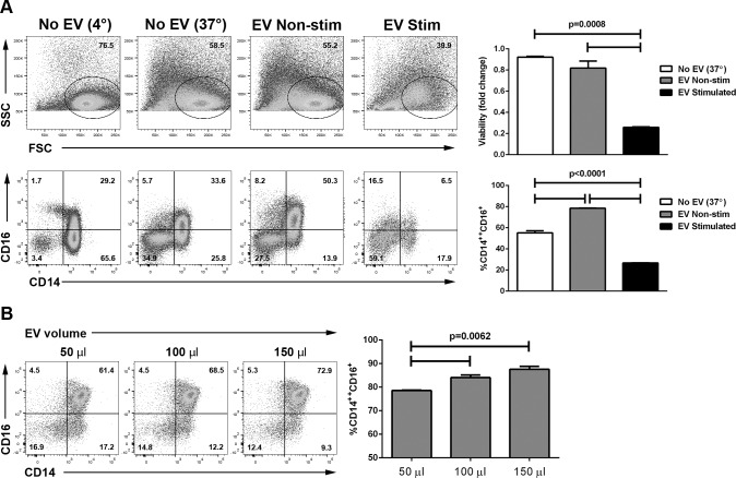
Figure 3.
Modulation of human monocyte phenotype by ARPE-19–derived EV. (A) Extracellular vesicles were isolated from ARPE-19 stimulated (EV: stimulated) or not (EV: nonstimulated) with IL-1β, IFN-γ, and TNF-α 10 ng/mL each for 48 hours, and 75 μL of EV isolate (1.1e9 ± 4.8e7 particles/mL for cytokine-stimulated ARPE-19 and 9.0e8 ± 1.6e7 particles/mL for nonstimulated ARPE-19) were cultured with elutriated human monocytes (0.5–1 × 10⁶ cells/well) for 24 hours. Cells were then stained with DAPI to assess viability and for CD14 and CD16 and analyzed by flow cytometry. Top row: dot plots showing forward (FSC) and side scatter (SSC); graph on the right shows the proportion of viable cells compared with those kept at 4° during the culture period. Bottom row: dot plots showing CD14 and CD16 expression; graph on the right shows the percentage of intermediate (CD14++CD16+) monocytes of all CD14+ cells in the different groups. Data representative of three independent experiments. (B) Extracellular vesicles were isolated from ARPE-19 not exposed to cytokines, and the indicated volume of EV isolate (2.2e8 ± 4.0e7 particles/mL) was cultured with elutriated human monocytes for 24 hours. Cells were then stained for CD14 and CD16 and analyzed by flow cytometry. * P < 0.05; P values calculated by ANOVA with Tukey's multiple comparisons test.

