
Fig. 2.
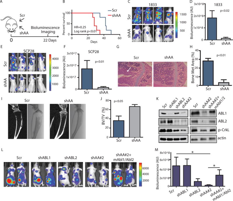
Knockdown of ABL kinases decreases breast cancer bone metastasis.
(A) The experimental design. (B) Survival of mice after intracardiac injection of 1833 (1×10⁵) breast cancer cells transduced with control shRNA (Scr) or shRNAs against ABL1 and ABL2 (shAA). N=10 mice/per group. (C–F) Bioluminescent images (C, E) of bone metastasis from representative mice at day 22 after inoculation with 1833 cells (N=10 mice/group) or day 35 after inoculation with SCP28 cells (N=8 mice/group). Quantification of bone metastases (D, F). (G–H) Representative HE staining (G) and quantification of HE-stained tumor area of bone lesions. Arrows indicate tumor. N=3 mice/group. Scale bar=200μM. Met, metastatic. (I–J) Representative Xray and μCT reconstruction (I) and quantification of bone volume/total volume from μCT analysis of the mouse tibias (J). N=3 mice/group. (K) Representative immunoblots of 1833 cells transfected with control shRNA (Scr), shRNA against ABL1 (shABL1), ABL2 (shABL2), and shRNA #2 against both ABL1 and ABL2 (shAA#2), and ABL1/ABL2 knockdown cells with overexpression of mouse Abl1/Abl2 (shAA+mAbl1/Abl2). N=3 blots. p, phosphorylated. (L) Bioluminescent images of bone metastases from representative mice at day 18 after inoculation. N=8 mice/group. (M) Quantification of (L). * p<0.05, calculated using One-Way ANOVA followed by Tukey’s HSD.