
Fig. 4.
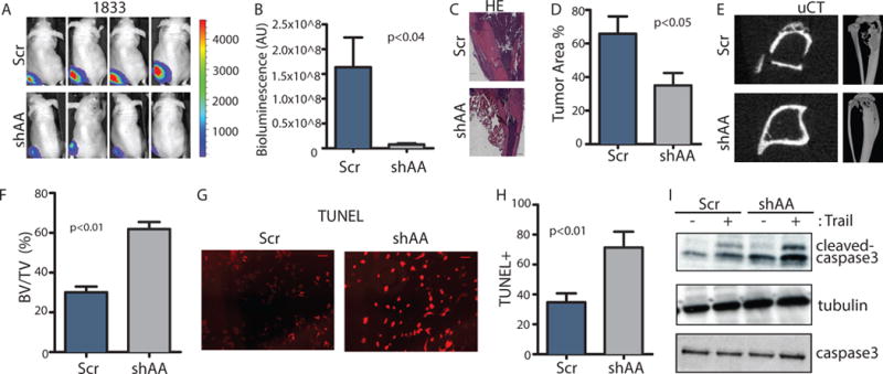
ABL kinases are required for tumor survival and tumor-induced osteolysis in the bone microenvironment.
(A–B) 1×10⁵ control or ABL1/ABL2 knockdown 1833 cells were injected directly into the tibias of the mice. Representative bioluminescent images (A) taken at day 21 after inoculation and quantification (B) of bone lesions are shown. N=5 mice/group. (C–D) Representative HE staining (C) and quantification (D) of HE-stained tumor area of mouse tibias from each group. N=3 mice/group. Scale bar=500μM. (E–F) Representative 3D μCT reconstruction of mouse tibias (E) and quantification (F) of bone volume/total volume (BV/TV) from μCT analysis. N=3 mice (G–H) Representative images (G) of TUNEL staining of cells treated with TRAIL and the indicated shRNA and quantification of the percent of TUNEL-positive cells (H). N=3 biological replicates. Scale bar=100μM. (I) Immunobloting was performed using the indicated antibodies on whole-cell lysates from cells incubated or not with TRAIL. N=3 blots.