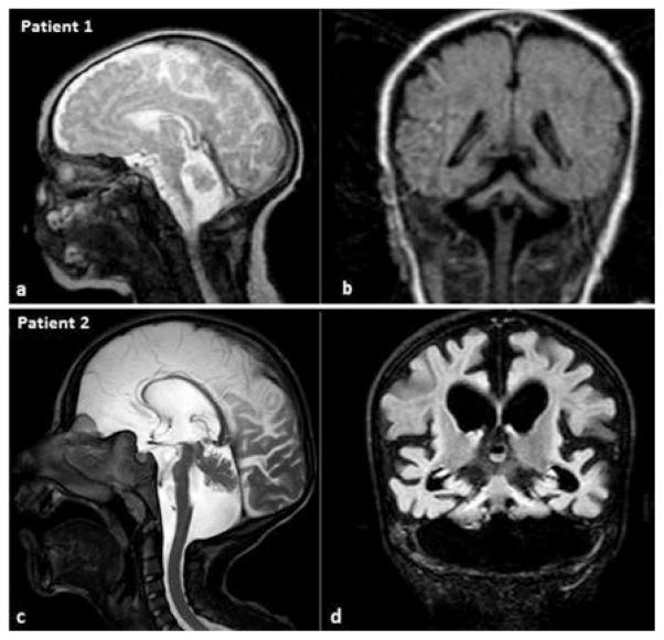
Fig. 1.
Cranial MRI images of patient 1 (a,b) during the neonatal period, and patient 2 (c,d) at the age of 8 years. Mid-sagittal T2-weighted (a,c) and coronal FLAIR (b,d) images show a flat ventral pons, cerebellar hypoplasia with the hemispheres being more affected and the vermis relatively spared (“dragonfly-like” pattern). In patient 2, cerebral atrophy is also present and cerebellar atrophy is more prominent, giving rise to an enlarged fourth ventricle.

