Fig. 2.

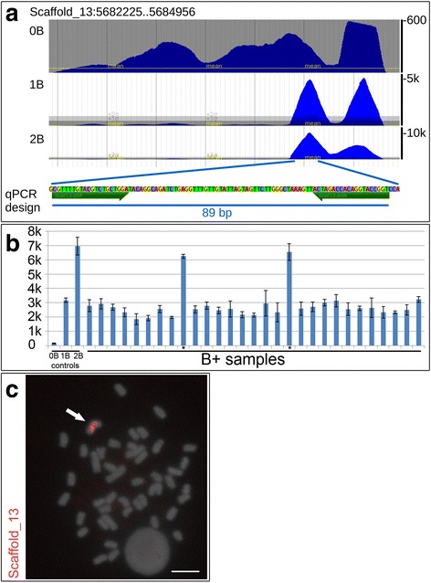
Development of and results observed for the qPCR marker analysis based on scaffold_13. a Scheme for primer design over a specific genomic region on scaffold_13 of 0B, 1B and 2B samples. Note the higher coverage for 1B (5,000× coverage) and 2B (10,000× coverage) samples compared with the 0B sample. b Graphic plot depicting the relative number of copies detected among B+ samples (1B and 2B) and control samples (0B). Note that 2B samples (asterisks) present approximately two-fold as many copies compared with the 1B samples. The standard deviation is presented for each sample. c FISH using the qPCR marker region sequence as a probe. An arrow indicates the B chromosome, and the scale bar is 5 μm
