
Figure 1.
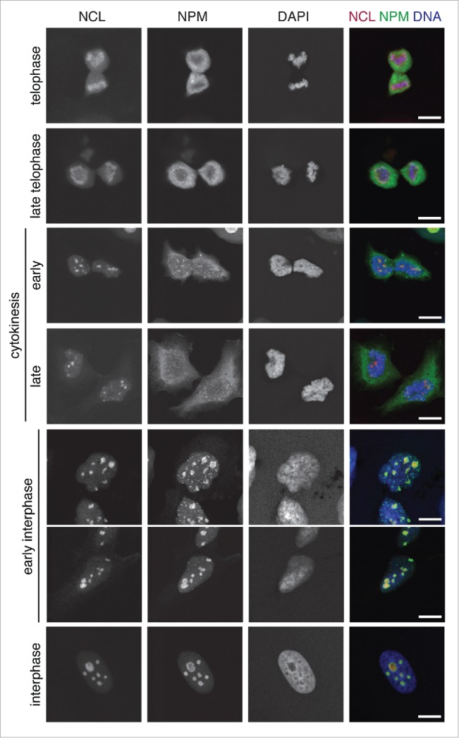
Cell cycle-dependent structural changes of the nucleolus. Confocal laser scanning microscopy (CLSM) images showing the nucleolar marker proteins nucleolin (NCL, red, stably expressed RFP-NCL) and nucleophosmin (NPM, green, immunofluorescence) with DNA (DAPI, blue) counterstaining in U2OS cells at different stages of the cell cycle. As evident from the NCL and NPM distribution, nucleoli are still completely disrupted during telophase and fully assembled during interphase. Scale bars, 10 µm.