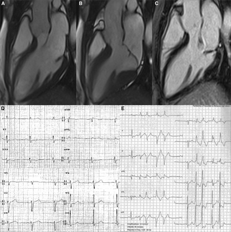
Figure 2.

Representative case of arrhythmic mitral valve prolapse with mitral annular disjunction, curling, and late gadolinium enhancement (LGE). A 36-y-old woman with mitral valve prolapse and complex ventricular arrhythmias. On cine cardiac magnetic resonance (CMR) 3-chamber, long-axis view (diastolic frame A, systolic frame B), a mitral annulus disjunction is detectable; on contrast-enhanced CMR, a midmural LGE in the LV inferobasal region under posterior valve leaflet is visible (C). The 12-lead ECG (D) shows a negative T wave in III-aVF. Nonsustained ventricular tachycardia with right bundle branch block morphology originating from the LV inferobasal wall near the mitral annulus is also recorded in the 24-h Holter ECG (E).
