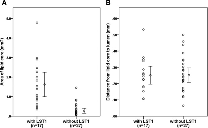
Figure 2.

A, Area of lipid core assessed by histological slides comparing low signal on T1-weighted fat-suppressed images (LST1) and without LST1 in the locations with lipid core. Lipid core area is significantly larger in the plaque with LST1 group than without LST1 group. B, Distance from lipid core to the lumen assessed by histological slides comparing LST1 and without LST1 in the locations with lipid core. Distance from lipid core to the lumen shows no difference in the plaque with LST1 group and without LST1 group.
