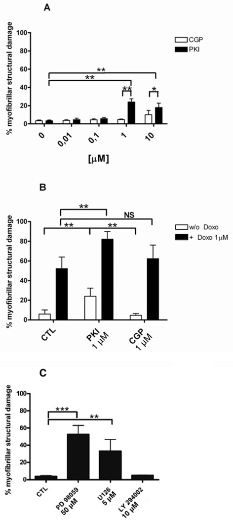
FIG. 3.
Quantification of myofibrillar structural damage. A: Comparison of different doses of PKI and CGP; B: PKI 1 µM and CGP 1 µM were combined with Doxo 1 µM (filled bars); C: Cardiomyocytes were treated with PD98059 50 µM, U126 5 µM or LY294002 10 µM. A–C: *p<0.01; **p<0.001; One-way ANOVA p<0.00001. N=6 independent experiments, 700 cells counted per condition.

