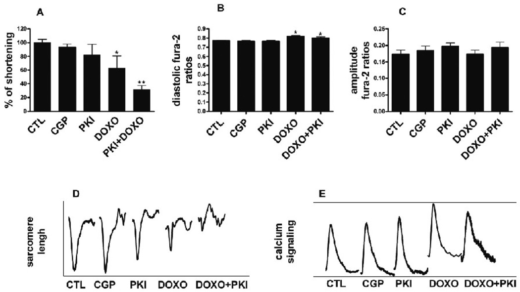
FIG. 8.
Excitation-contraction coupling. Cardiomyocytes cultured for 10 days were treated for 48h with PKI 1 µM, CGP 1 µM, Doxo 1 µM, or PKI 1 µM in combination with Doxo 1 µM. Sarcomere shortening and calcium transients were determined in 2 Hz paced cells, as described in methods. Tracings (D and E) represent the average contraction and calcium transients of 39 cells (n=3, 13 cells for experiment). Results were normalized for the averaged values measured in untreated cells. A. Fractional shortening. *p<0.01 vs. CTL; **p<0.001 vs. CTL; One-way ANOVA p<0.00001. B. Diastolic fura-2 ratios. *p<0.01 vs. CTL; One-way ANOVA p<0.00001. C. Calcium amplitude D. Averaged contraction transients. E. Averaged calcium transients.

