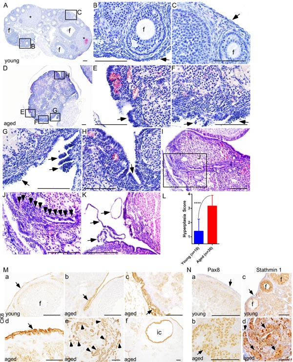
Figure 2. Deregulated growth of ovarian surface epithelium in aged mouse ovaries.

A.-C. Histological analysis of a young mouse ovary (8 wk) showing follicles (B; marked with f), corpora lutea (asterisks in panel A), and a single flattened layer of ovarian surface epithelial cells (C; arrow). D. An abnormal looking hyperplastic ovary of aged mice. Higher magnification images of boxed areas in panel D revealed irregular morphology and ovarian surface epithelial cell hyperplasia (arrows in E. and F.), epithelial multilayering and abnormal papillary growth (arrows in G.), and epithelial invaginations into stroma H. I. and J. OSE cells in an aged ovary infiltrating (arrowheads in panel J) into the stroma forming inclusion cysts and glandular structures. Panel J is a higher magnification picture of boxed area in panel I. Extensive epithelial shedding and outgrowths (arrows in panel K.) in aged ovaries. L. Significant increase in ovarian surface epithelial hyperplasia score of ovaries of the aged mice compared to young controls. M. Epithelial origin of abnormal lesions confirmed by CK8 staining. (M., a) Normal CK8-postive OSE cells (arrow) were seen in young mouse ovary. (M., b-f) CK8 expression was observed in abnormal invasive and hyperplastic growths (arrow in panel b-d, arrowheads in panel e) of OSE in aged mice. (M., f) CK8-positive inclusion cyst (ic) present in an aged ovary. (N., b and d) PAX8 and Stathmin 1 expression in invasive and hyperplastic epithelial growths (arrow) of aged mice ovaries. (N., a and c) Ovarian surface epithelium (arrow) of young mouse ovary was negative for PAX8 and Stathmin 1 staining. (N., c) Stathmin 1 was expressed by the somatic cells of follicles (marked with f) in young ovaries. Bars: 100 μm.
