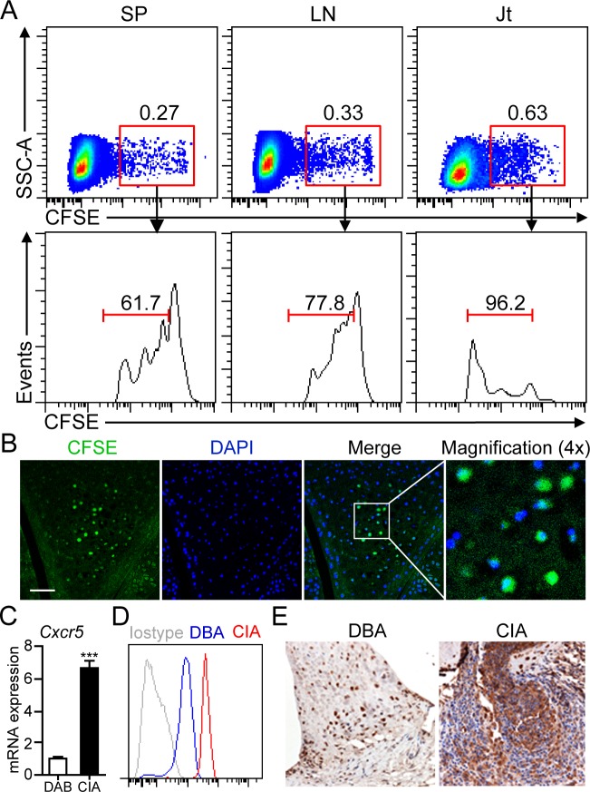
Figure 2. B1a cells migrate from PC to the joint tissue of CIA mice.
A. Sorting-purified peritoneal B1a cells were stained with CFSE and intraperitoneally transferred into DBA mice and followed by CII immunization for CIA induction. On day 17 after cell transfer, CFSE+ B1a cells in cell suspensions from the spleen (SP), draining lymph nodes (LN) and joint tissues (Jt) were detected by flow cytometry. Flow profiles are representative from three independent experiments. B. CFSE+ B1a cells accumulated in the synovium of knee joint of B1a-transferred CIA mice were detected by confocal microscopy (n = 5). Scale bar, 50 μm. C., D. CXCR5 expression on peritoneal B1a cells from DBA and CIA (14 dpi) mice were measured by q-PCR in C and flow cytometry in D (n = 6). Data in C were shown as mean ± SD (***, p < 0.001). E. CXCL13 expression in the synovium of knee joints of DBA and CIA mice on 17 dpi were measured by immunohistochemistry (IHC) staining. Nucleus was stained with hematoxylin solution. CXCL13-expressing cells are stained an intense brown (Original magnification, ×100) (n = 5).

