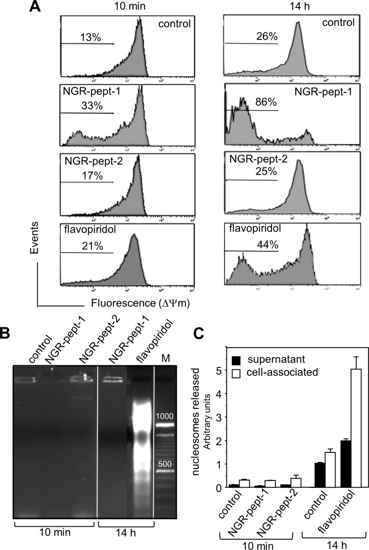
Figure 5. NGR-peptide-1 induces mitochondrial membrane depolarization in the absence of DNA fragmentation.
(A–C) U937 cells were cultured for 10 min and/or 14 h in the absence or presence of 50 μM NGR-peptides or 100 nM flavopiridol (positive control). (A) Then, cells were labelled with the fluorescent probe JC-1. The loss of mitochondrial membrane potential (ΔΨm) is characterized by a significant shift from red (polarization) fluorescence to green (depolarization) fluorescence. Diluted DMSO (corresponding to 100 nM flavopiridol) had no effect on ΔΨm. The percentages refer to ΔΨm dissipation. DNA fragmentation was evaluated by (B) the detection of an oligonucleosome ladder by agarose gel electrophoresis and (C) release of histone-associated DNA fragments (mono- and oligonucleosomes). Data are mean ± SD of three separate determinations.

