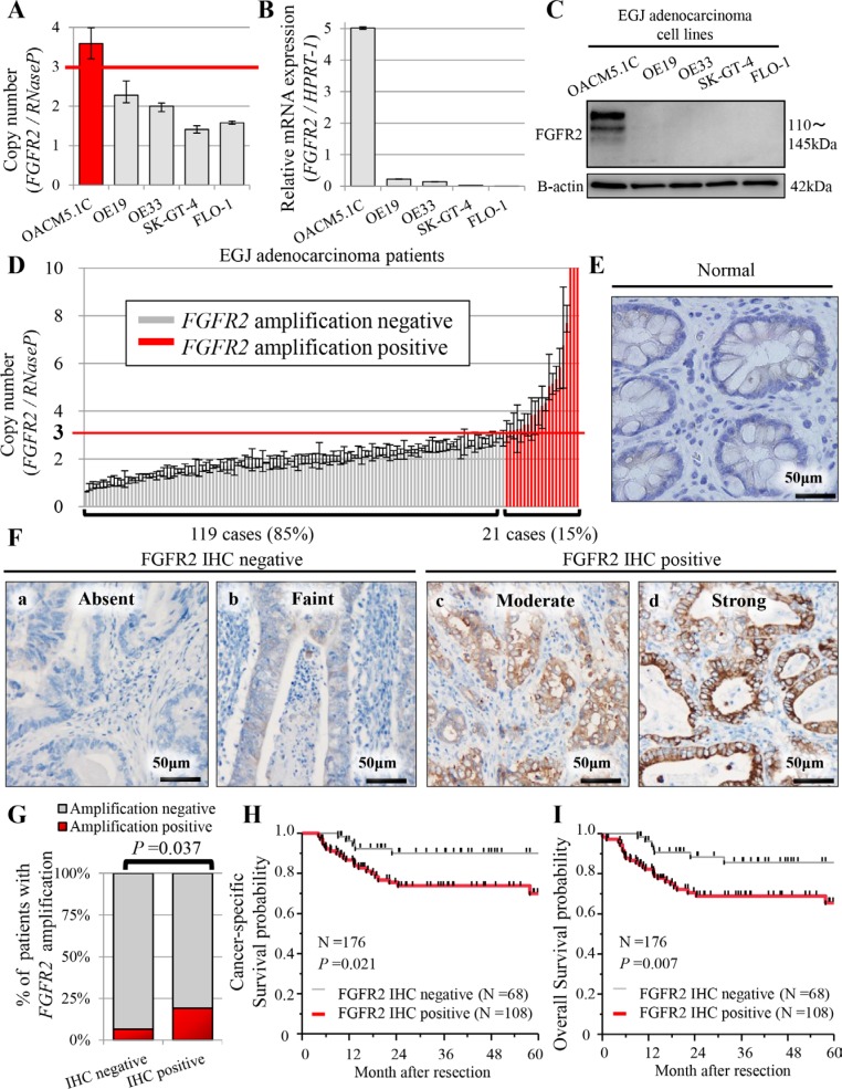
Figure 1. Profiles of FGFR2 status in five human EGJ adenocarcinoma cell lines (A–C), and in patients with EGJ adenocarcinoma (N = 176) (D–I).
(A) FGFR2 copy number obtained in real-time PCR assay; (B) mRNA expression by qRT-PCR assay; (C) FGFR2 expression by western blot analysis; (D) Distributions of FGFR2 copy number (N = 140). For FGFR2 amplification, the copy number gain must exceed 3.0 copies. FGFR2 amplification was observed in 21 cases (21/140 = 15%); (E) FGFR2 was not expressed in normal glandular epithelium; (F) (a, b) Cases with absent or faint FGFR2 staining were assessed as FGFR2 IHC-negative; (c, d) cases with moderate or strong FGFR2 staining were FGFR2 IHC-positive; (G) Association between FGFR2 IHC positivity and FGFR2 amplification; (H) Cancer-specific survival in positive and negative FGFR2 IHC cases; (I) Overall survival in positive and negative FGFR2 IHC cases.

